Current issue
Online first
Archive
About the Journal
Editorial Office
Editorial Board
Copy right and self-archiving policy
Peer review process
Instructions for Reviewers
Printed version subscription
Abstracting and indexing
Contact
Instructions for Authors
Policies
General information
Open Access, Licensing terms, Commercial use and Copyright terms policies
Self-archiving policy and Archive policies
Article Correction and Withdrawal policy
Manuscript Submission policy
Authorship policy
Conflict of Interest policy
Language considerations policy
Plagiarism and Duplicate publications policy
Ethics policy
Review process policy
Acceptance of manuscripts policy
Online First Articles and Special Issues policies
Generative artificial intelligence (AI) policy
Advertising policy
Article publication charges
Policies
General information
Open Access, Licensing terms, Commercial use and Copyright terms policies
Self-archiving policy and Archive policies
Article Correction and Withdrawal policy
Manuscript Submission policy
Authorship policy
Conflict of Interest policy
Language considerations policy
Plagiarism and Duplicate publications policy
Ethics policy
Review process policy
Acceptance of manuscripts policy
Online First Articles and Special Issues policies
Generative artificial intelligence (AI) policy
Advertising policy


ORIGINAL PAPER
Kidney stone formation in exocrine pancreatic insufficient pigs fed an oxalate enriched diet
1
The Kielanowski Institute of Animal Physiology and Nutrition, Polish Academy of Sciences, Department of Animal Physiology, 05-110 Jabłonna, Poland
2
The Kielanowski Institute of Animal Physiology and Nutrition, Polish Academy of Sciences, Large Animal Models Laboratory, 05-110 Jabłonna, Poland
3
Research Institute of Animal Husbandry, 32-083 Balice, Poland
4
Anara AB, 23132 Trelleborg, Sweden
5
Institute of Clinical Dentistry, Fondazione Policlinico Universitario A. Gemelli IRCCS, Universita Cattolica del Sacro Cuore, Oral Surgery and Implantology Unit, Division of Oral Surgery and Implantology, Department of Head and Neck, 00168 Rome, Italy
6
Collegium Medicum Nicolaus Copernicus University, Department of Interventional Dentistry, 85-067 Bydgoszcz, Poland
7
Department of Biology, Lund University, Lund, Sweden
8
Institute of Rural Health, Department of Medical Biology, 20-950 Lublin, Poland
Publication date: 2026-05-22
Corresponding author
K. Zaworski
The Kielanowski Institute of Animal Physiology and Nutrition, Polish Academy of Sciences, Department of Animal Physiology, 05-110 Jabłonna, Poland
The Kielanowski Institute of Animal Physiology and Nutrition, Polish Academy of Sciences, Department of Animal Physiology, 05-110 Jabłonna, Poland
J. Anim. Feed Sci. 2026;35(3):e36
KEYWORDS
calcium oxalate nephrolithiasisexocrine pancreatic insufficiencyhyperoxaluriakidney stonespancreatic enzyme replacement therapy
TOPICS
ABSTRACT
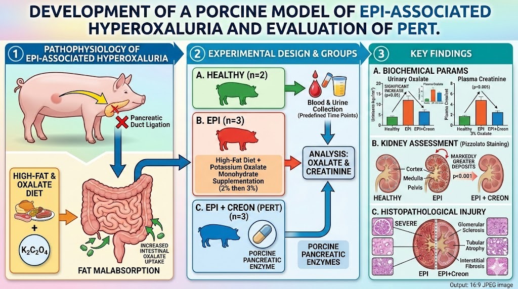
Exocrine pancreatic insufficiency (EPI) causes fat malabsorption, which can increase intestinal oxalate uptake and promote calcium oxalate kidney stone formation. The aim of this study was to develop a porcine model of EPI-associated hyperoxaluria and nephrolithiasis, and to evaluate whether pancreatic enzyme replacement therapy (PERT; Creon®) reduces renal injury. A randomised controlled experiment included eight pigs: six underwent pancreatic duct ligation to induce EPI and were allocated to the EPI group (n = 3) or the EPI+Creon group (n = 3), while two healthy pigs served as comparators. All pigs were fed a high-fat diet; EPI groups additionally received potassium oxalate monohydrate supplementation (2%, followed by 3%). The EPI+Creon group was administered porcine pancreatic enzymes. Blood and urine samples were collected at predefined time points for measurement of oxalate and creatinine levels. At study termination, kidneys were assessed for calcium oxalate deposition using Pizzolato staining and for histopathological damage using semi-quantitative scoring. Oxalate supplementation significantly increased urinary and plasma oxalate concentrations in pigs with EPI (P < 0.05) and was associated with higher plasma creatinine at the 3% oxalate dose (P = 0.005). Calcium oxalate deposits were markedly more abundant in the renal cortex, medulla, and pelvis in EPI pigs than in EPI+Creon and healthy animals (P < 0.001). Severe glomerular sclerosis, tubular atrophy, and interstitial fibrosis were observed in EPI pigs and were only partially attenuated by PERT. In conclusion, oxalate-fed pigs with EPI develop hyperoxaluria, nephrolithiasis, and renal injury. PERT reduces but does not fully prevent these biochemical and structural kidney abnormalities.
FUNDING
This research was funded by Anara AB.
CONFLICT OF INTEREST
The authors declare that there is no conflicts of interest.
REFERENCES (35)
1.
Aris R., Lester G., Dingman S., Ontjes D.A., 1999. Altered calcium homeostasis in adults with cystic fibrosis. Osteoporos. Int. 10, 102–108, https://doi.org/10.1007/s00198....
2.
Ashry M., Galal El-Sahra D., Gaber D.A., Mustafa A.M., Abdel-Wahhab K.G., 2021. Nephroprotective effect of costus (Saussurea costus) ethanolic extract on oxaliplatin®-induced nephrotoxicity in adult male Wistar rats. Pak. J. Biol. Sci. 24, 830–839, https://doi.org/10.3923/pjbs.2....
3.
Asplin J.R., 2002. Hyperoxaluric calcium nephrolithiasis. Endocrinol. Metab. Clin. North Am. 31, 927–949, https://doi.org/10.1016/S0889-....
4.
Belostotsky R., Seboun E., Idelson G.H., Milliner D.S., Becker-Cohen R., Rinat C., Shalev H., Raas-Rothschild A., 2010. Mutations in DHDPSL are responsible for primary hyperoxaluria type III. Am. J. Hum. Genet. 87, 392–399, https://doi.org/10.1016/j.ajhg....
5.
Bhasin B., Ürekli H.M., Atta M.G., 2015. Primary and secondary hyperoxaluria: Understanding the enigma. World J. Nephrol. 4, 235–244, https://doi.org/10.5527/wjn.v4....
6.
Cochat P., Liutkus A., Fargue S., Basmaison O., Ranchin B., Rolland M.O., 2006. Primary hyperoxaluria type 1: still challenging! Pediatr. Nephrol. 21, 1075–1081, https://doi.org/10.1007/s00467....
7.
Cochat P., Rumsby G., 2013. Primary hyperoxaluria. N. Engl. J. Med. 369, 649–658, https://doi.org/10.1056/NEJMra....
8.
Cregeen D.P., Rumsby G., 1999. Recent developments in our understanding of primary hyperoxaluria type 2. J. Am. Soc. Nephrol. 10, 348–350.
9.
Danpure C.J., 2005. Molecular etiology of primary hyperoxaluria type I: New directions for treatment. Am. J. Nephrol. 25, 303–310, https://doi.org/10.1159/000087....
10.
Directive 2010/63/EU of the European Parliament and of the Council of 22 September 2010 on the protection of animals used for scientific purposes. Official Journal of the European Union L 276, 20.10.2010, p. 33–79
11.
Fischer A.H., Jacobson K.A., Róża J., Zeller R., 2008. Paraffin embedding tissue samples for sectioning. Cold Spring Harb. Protoc. 2008, 49, https://doi.org/10.1101/pdb.pr....
12.
Glew R.H., Sun Y., Horowitz B.L., Konstantinov K.N., Barry M., Fair J.R., Massie L., Tzamaloukas A.H., 2014. Nephropathy in dietary hyperoxaluria: A potentially preventable acute or chronic kidney disease. World J. Nephrol. 3, 122–142, https://doi.org/10.5527/wjn.v3....
13.
Harambat J., Fargue S., Acquaviva C., et al., 2010. Genotype-phenotype correlation in primary hyperoxaluria type 1: the p.Gly170Arg AGXT mutation is associated with a better outcome. Kidney Int. 77, 443–449, https://doi.org/10.1038/ki.200....
14.
Hatch M., Freel R.W., 2008. The roles and mechanisms of intestinal oxalate transport in oxalate homeostasis. Semin. Nephrol. 28, 143–151, https://doi.org/10.1016/j.semn....
15.
Higgins J.P., 2023. Ten traits of great physicians! And tips to help you improve. Am. J. Med. 136, 355–359, https://doi.org/10.1016/j.amjm....
16.
Holmes R.P., Goodman H.O., Assimos D.G., 2001. Contribution of dietary oxalate to urinary oxalate excretion. Kidney Int. 59, 270–276, https://doi.org/10.1046/j.1523....
17.
Hoppe B., 2012. An update on primary hyperoxaluria. Nat. Rev. Nephrol. 8, 467–475, https://doi.org/10.1038/nrneph....
18.
Hoppe B., Bec B.B., Milliner D.S., 2009. The primary hyperoxalurias. Kidney Int. 75, 1264–1271, https://doi.org/10.1038/ki.200....
19.
Hoppe B., Kemper M.J., Hvizd M.G., Sailer D.E., Langman C.B., 1998. Simultaneous determination of oxalate, citrate and sulfate in children’s plasma with ion chromatography. Kidney Int. 53, 1348–1352, https://doi.org/10.1046/j.1523....
20.
Hoppe B., Latta K., von Schnakenburg C., Kemper M.J., 2005. Primary hyperoxaluria—the German experience. Am. J. Nephrol. 25, 276–281, https://doi.org/10.1159/000086....
21.
Kumar R., Ghoshal U.C., Singh G., Mittal R.D., 2004. Infrequency of colonization with Oxalobacter formigenes in inflammatory bowel disease: possible role in renal stone formation. J. Gastroenterol. Hepatol. 19, 1403–1409, https://doi.org/10.1111/j.1440....
22.
Liebman M., Al-Wahsh I.A., 2011. Probiotics and other key determinants of dietary oxalate absorption. Adv. Nutr. 2, 254–260, https://doi.org/10.3945/an.111....
23.
Lieske J.C., Tremaine W.J., De Simone C., O’Connor H.M., Li X., Bergstralh E.J., Goldfarb D.S., 2010. Diet, but not oral probiotics, effectively reduces urinary oxalate excretion and calcium oxalate supersaturation. Kidney Int. 78, 1178–1185, https://doi.org/10.1038/ki.201....
24.
Lunney J.K., 2007. Advances in swine biomedical model genomics. Int. J. Biol. Sci. 3, 179–184, https://doi.org/10.7150/ijbs.3....
25.
Mitchell T., Kumar P., Reddy T., Wood K.D., Knight J., Assimos D.G., Holmes R.P., 2019. Dietary oxalate and kidney stone formation. Am. J. Physiol. Renal Physiol. 316, 409-413, https://doi.org/10.1152/ajpren....
26.
Nayir Buyuksahin H., Emiralioglu N., Ademhan Tural D., et al., 2023. Coexistence of cystic fibrosis with other genetic disorders: A case series. Pediatr. Pulmonol. 58, 345–347, https://doi.org/10.1002/ppul.2....
27.
Ozçelik U., Beşbaş N., Göçmen A., Akata D., Akhan O., Ozgüç M., Kiper N., 2004. Hypercalciuria and nephrocalcinosis in cystic fibrosis patients. Turk. J. Pediatr. 46, 22–27.
28.
Pierzynowski S., Swieboda P., Filip R., et al., 2012. Behavioral changes in response to feeding pancreatic-like enzymes to exocrine pancreatic insufficiency pigs. J. Anim. Sci. 90, 439–441, https://doi.org/10.2527/jas.53....
29.
Pizzolato P., 1964. Histochemical recognition of calcium oxalateJournal of Histochemistry and Cytochemistry, 12, 333–336, https://doi.org/10.1177/12.5.3....
30.
Putman M.S., Anabtawi A., Le T., Tangpricha V., Sermet-Gaudelus I., 2019. Cystic fibrosis bone disease treatment: Current knowledge and future directions. J. Cyst. Fibros. 18, 56–65, https://doi.org/10.1016/j.jcf.....
31.
Siener R., Bangen U., Sidhu H., Hönow R., von Unruh G., Hesse A., 2013. The role of Oxalobacter formigenes colonization in calcium oxalate stone disease. Kidney Int. 83, 1144–1149, https://doi.org/10.1038/ki.201....
32.
Siener R., Ernsten C., Speller J., Scheurlen C., Sauerbruch T., Hesse A., 2024. Intestinal oxalate absorption, enteric hyperoxaluria, and risk of urinary stone formation in patients with Crohn’s disease. Nutrients 16, 264, https://doi.org/10.3390/nu1602....
33.
Swindle M.M., Makin A., Herron A.J., Clubb F.J. Jr., Frazier K.S., 2012. Swine as models in biomedical research and toxicology testing. Vet. Pathol. 49, 344–356, https://doi.org/10.1177/030098....
34.
Szkopek D., Pierzynowski S.G., Pierzynowska K., et al., 2024. A review: Pancreatic enzymes in the treatment of chronic pancreatic insufficiency in companion animals. J. Vet. Intern. Med. 38, 2026–2033, https://doi.org/10.1111/jvim.1....
35.
Zaworski K., Wychowański P., Szkopek D., Woliński J., Donaldson J., Pierzynowski S., Pierzynowska K., 2025. The regulatory role of pancreatic enzymes in the maintenance of small intestinal structure and enterocyte turnover with special reference to alpha amylase. Int. J. Mol. Sci. 26, 249, https://doi.org/10.3390/ijms26....
| ISSN: | 1230-1388 |
Assigning DOI numbers, introducing articles from supplements and recent publications to POL-index database, maintaining anti-plagiarism detection, electronic system to proceed manuscripts and webpage of the Journal with interactive pdf files, and language correction of manuscripts published in Journal of Animal and Feed Sciences are financed in the years 2018–2019 by the Ministry of Science and Higher Education from the funds for science popularization activities, Agreement No. 631/P-DUN/2018.

© 2006-2026 Journal hosting platform by Bentus
We process personal data collected when visiting the website. The function of obtaining information about users and their behavior is carried out by voluntarily entered information in forms and saving cookies in end devices. Data, including cookies, are used to provide services, improve the user experience and to analyze the traffic in accordance with the Privacy policy. Data are also collected and processed by Google Analytics tool (more).
You can change cookies settings in your browser. Restricted use of cookies in the browser configuration may affect some functionalities of the website.
You can change cookies settings in your browser. Restricted use of cookies in the browser configuration may affect some functionalities of the website.